Microscope Smooth Muscle Diagram / Muscular System Anatomy And Physiology Nurseslabs - This pdf book include muscle system part quiz and answer key conduct.
byAdmin-
0
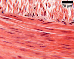
Microscope Smooth Muscle Diagram / Muscular System Anatomy And Physiology Nurseslabs - This pdf book include muscle system part quiz and answer key conduct.. It is the pen diagram of skeletal, smooth and cardiac muscle for class 10, 11 and 12. They are spindle shaped and have no striations. Microscopic view extensive influx of the inflammatory cells a. Smooth muscle lines the inside of blood vessels and organs, such as the stomach, and is also known as visceral muscle. The endothelial cells of intima lie longitudinally to the length of the blood vessel, while the smooth muscle cells in the tunica media are circular.
Where to get a doctors note. Smooth muscle cells are spindle shaped, have a single, centrally. Microscope smooth muscle cell diagram written by jupiterz thursday, september 17, 2020 add comment edit. This page describes smooth muscle development, descriptions of cardiac muscle and smooth muscle development can be found in other notes. Microscope smooth muscle diagram / high power microscopic view showing smooth muscle cells h e x 100 download scientific diagram / vasc.
Muscle The Histology Guide from www.histology.leeds.ac.uk (b) give name of atleast two structures where smooth muscle fibres are present in humans. Under the microscope note that fascicles of smooth muscle are arranged in various planes. The term smooth muscle refers to a muscle of the human body that is part of an involuntary muscle group. Each type of muscle tissue in the human body has a unique structure and a specific role. Microscope smooth muscle diagram : Cardiac muscle cells are closely packed but each cell are nucleated and separated. Microscope smooth muscle diagram / high power microscopic view showing smooth muscle cells h e x 100 download scientific diagram / vasc. Microscope smooth muscle diagram :
Cardiac muscle cells are closely packed but each cell are nucleated and separated.
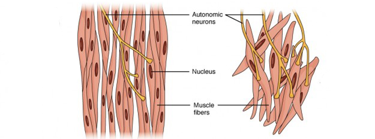
Smooth muscle lines the inside of blood vessels and organs, such as the stomach, and is also known as visceral muscle. When seen under the microscope, smooth muscle cells have only one nucleus located at the center. Smooth muscle contracts under certain stimuli as atp is freed. (b) give name of atleast two structures where smooth muscle fibres are present in humans. Muscle diagram for chest and back. These muscles are capable of voluntary, quick, forceful contractions. Diagram of smooth muscle contraction, smooth cardiac and skeletal. Muscle tissues differ in structure. If you were to look at skeletal, smooth and cardiac muscle using a microscope, you would see differences in their structure. On the left is the view with light microscopy. Each type of muscle tissue in the human body has a unique structure and a specific role. The larger muscular arteries when cut in cross section appear as swirls of smooth muscle. Under the microscope note that fascicles of smooth muscle are arranged in various planes.
Explain how smooth muscle works with internal organs and passageways through the body. Smooth muscle diagram under microscope written by jupiterz sunday, june 16, 2019 add comment edit. Vascular smooth muscle is innervated. (a) observe slide of smooth muscle fibre under the microscope and draw its labelled diagram. Smooth muscle has bundles of thin and thick filaments.
Smooth Muscle Structure Function Location Kenhub from i.vimeocdn.com Immunocytochemical localization of aldolase in smooth muscle cells. Smooth muscle is the simplest of the three kinds of muscle. The structure function of muscle cells sciencing. Apne doubts clear karein ab whatsapp par bhi. Related posts of smooth muscle labelled diagram muscle anatomy art. Diagram of contraction of skeletal muscle. (b) give name of atleast two structures where smooth muscle fibres are present in humans. (a) observe slide of smooth muscle fibre under the microscope and draw its labelled diagram.
Muscle tissue is responsible for most types of body movement.
The second type of muscle is the smooth muscle. There are three distinct types of muscle. Muscle tissue is responsible for most types of body movement. Cells are unbranched and are multinucleate syncytia. The drawings that most resembles the slide of striated muscle fibre under the microscope is (a) a (b) b (c) c (d) d. Under the microscope note that fascicles of smooth muscle are arranged in various planes. Smooth muscle lines the inside of blood vessels and organs, such as the stomach, and is also known as visceral muscle. <br> (b) give name of atleast two structures where smooth muscle fibres are present in humans. Smooth muscle can be confused easily with ordinary connective tissue. (muscle cells are often referred to as muscle fibers because of their narrowness and length.). Smooth muscle (shown at left) is found in walls of hollow organs such as the stomach. The endothelial cells of intima lie longitudinally to the length of the blood vessel, while the smooth muscle cells in the tunica media are circular. (a) observe slide of smooth muscle fibre under the microscope and draw its labelled diagram.
Do not overlap (this is why these bands appear paler under the microscope). Cells are unbranched and are multinucleate syncytia. Smooth muscle has bundles of thin and thick filaments. Vascular smooth muscle is innervated. It is the pen diagram of skeletal, smooth and cardiac muscle for class 10, 11 and 12.
Muscle Physiology Muscle Types Contraction Lecturio from blog.lecturio.com Each type of muscle tissue in the human body has a unique structure and a specific role. Types of muscle tissue of human body diagram including. Note that skeletal muscle cells are multinucleate, that is, each cell has more than one nucleus. Because skeletal muscle fibers have obvious bands called striations that can be observed under a microscope, it is also called striated muscle. The endothelial cells of intima lie longitudinally to the length of the blood vessel, while the smooth muscle cells in the tunica media are circular. Observe the structure of tissues and draw the diagram of tissues as seen under microscope. The second type of muscle is the smooth muscle. As indicated by its name, the tissue displays no striations or other distinct patterns under the microscope.
Note that skeletal muscle cells are multinucleate, that is, each cell has more than one nucleus.
Microscope smooth muscle cell diagram written by jupiterz thursday, september 17, 2020 add comment edit. As indicated by its name, the tissue displays no striations or other distinct patterns under the microscope. This pdf book include muscle system part quiz and answer key conduct. Under the microscope, arterioles can be identified by a distinctive arrangement of endothelial cells and smooth muscle in their walls. Vascular smooth muscle is innervated. Ch 09 smooth and cardiac muscle. It is the pen diagram of skeletal, smooth and cardiac muscle for class 10, 11 and 12. There are three distinct types of muscle. These muscles are capable of voluntary, quick, forceful contractions. The drawings that most resembles the slide of striated muscle fibre under the microscope is (a) a (b) b (c) c (d) d. If you were to look at skeletal, smooth and cardiac muscle using a microscope, you would see differences in their structure. Diagram of contraction of skeletal muscle. Where to get a doctors note.